DIAART: a diaphragm implant for congenital diaphragmatic hernia
Congenital diaphragmatic hernia (CDH) is a severe genetic malformation caused by abnormal development of the diaphragm. It leads to abdominal organs herniating into the thoracic cavity, preventing normal lung development in utero. This condition affects 1 in 3,500 births, or around 50,000 newborns per year worldwide.
Researchers from INSERM (Unit 1121 – Biomaterials and Bioengineering), ICS, and engineers from Cetim Grand Est, all members of Carnot MICA, in close collaboration with pediatric surgeon Isabelle Talon, have developed an elastic material capable of adapting to the child’s growth. In the long term, this material could revolutionize the implants used to repair the diaphragm.
Current treatment and its limitations
The current treatment for newborns with CDH involves reducing the herniated organs and implanting a prosthesis made of polytetrafluoroethylene (PTFE). The goal is to close the diaphragm and prevent organs from moving back into the thoracic cavity.
However, this material is not elastic. Over time, it can lead to growth-related complications or even implant detachment, which may cause recurrence of the hernia.
The DIAART project: toward a new generation of implants
Through the DIAART resource recovery project funded by Carnot MICA, researchers aim to develop a biocompatible, elastic material that can grow in harmony with the child’s body.
This innovation could enable the creation of a new generation of diaphragmatic implants that adapt to physiological growth, significantly improving long-term outcomes for children born with CDH.
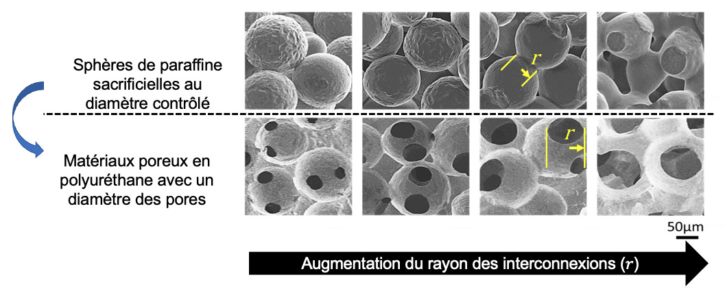
Development of a method to generate porous polyurethane materials with controlled architecture
The objective is to study how material structure influences the ability of different types of cells to colonize the scaffold. In this work, paraffin spheres with a rationalized size distribution are first compacted, heated, and then coated with polyurethane (top images). After dissolution of the spheres (bottom images), a material is obtained that features both porosity and a controlled size of the interconnections (r) between pores.
This approach makes it possible to create model systems to investigate how pore architecture affects cellular behavior.
From the outset, the researchers identified three key challenges to overcome: the material had to be biocompatible, elastic to follow the child’s growth, and porous to allow colonization by striated muscle cells similar to those of the diaphragm, while also remaining smooth to prevent adhesion of abdominal organs.
To address these challenges, the teams chose to develop a polyurethane-based film, a clinically approved material already known for its biocompatibility and elastic properties.
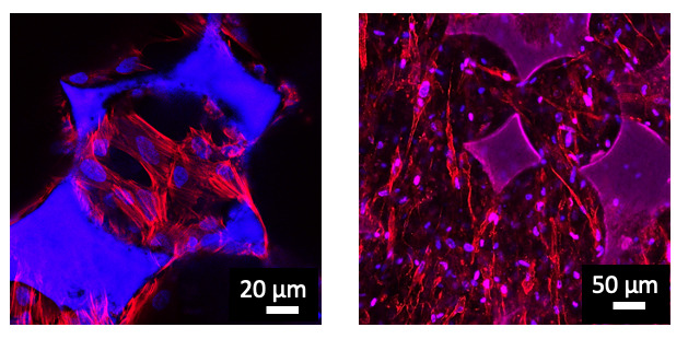
Study of cell colonization within porous materials
The two images correspond to materials in which pore diameters and interconnections were optimized to enable nearly complete cell colonization. Two different cell types were seeded into these structures: mesenchymal stem cells (left image) and fibroblasts (right image). This demonstrates the versatility of these systems for applications in tissue engineering.
After overcoming the first two major challenges, the remaining task was to develop a technique capable of introducing the required porosity for biological colonization. To achieve this, the researchers used sacrificial paraffin beads with controlled sizes and interconnections to create an optimal physical architecture for cell growth. Once this “template bed” was formed, it was coated with polyurethane, polymerized, and then immersed in a solvent to dissolve the beads and recover the porous film. A plasma and PDA treatment was then applied to remove the material’s hydrophobicity, which would otherwise prevent cell colonization in biological fluids.
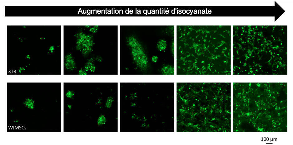
Once the film was produced, the researchers could study and optimize it to identify the best composition for an effective prosthesis. They discovered that the isocyanate index had a significant influence on cell adhesion, due to the microstructure of the molecule. The higher the index, the stronger the cell adhesion to the prosthesis. Additional treatments were also applied to enhance cell attachment, such as albumin incorporation, a low-cost natural protein.
Today, based on the full set of results and a comprehensive physicochemical analysis of the newly developed prosthesis, the researchers have validated an optimal composition to address congenital diaphragmatic hernia, paving the way for the development of other implants requiring similar properties.
However, one challenge remains: the material’s lack of resistance at suture points. This issue is now the focus of a new research project between the INSERM unit and the Institute of Chemistry and Processes for Energy, Environment and Health (ICPEES) in Strasbourg. The goal is to fabricate the material using spin coating, a technique for forming a thin, uniform layer by depositing a solution of the film material onto a flat substrate rotating at high speed.