ELECTRATPULP: revolutionizing endodontic care
ELECTRATPULP: revolutionizing endodontic care through tissue regeneration
With the ELECTRATPULP project, researchers from INSERM, ICPEES, IREPA LASER, and the “Autoimmunity, Transplantation, and Inflammation” laboratory aim to propose a new endodontic treatment method based on tissue regeneration. A key advantage of this approach is that it preserves the tooth’s biological response capacity while reducing intervention costs in cases of pulp tissue loss.
Introducing tissue regeneration into endodontic care
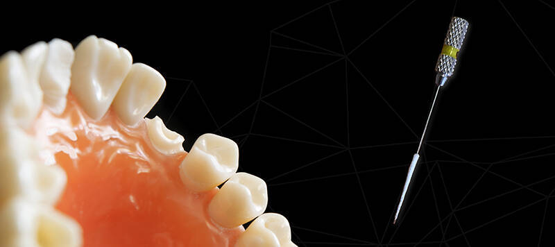
Despite numerous awareness campaigns, dental caries remain a major public health issue and still frequently lead to damage of the dental pulp tissue. The current endodontic treatment consists of three main steps:
- Removal of infected or necrotic tissue
- Cleaning of the root canal system
- Filling of the canal with thermoplastic materials such as gutta-percha
With more than 4 million procedures performed annually (HAS, 2004), treatment failures remain significant. Moreover, this approach has a major drawback: it eliminates the tooth’s biological integrity, leading to long-term weakening and eventual tooth loss, as the tooth can no longer detect bacterial attacks.
A regenerative alternative
The approach developed by the teams promotes the reconstruction of pulp tissue using the patient’s own stem cells. This “cell-homing” strategy offers several advantages:
- Reduced treatment costs
- Faster tissue development
- Guaranteed biocompatibility of the newly formed tissue
However, a major challenge remained: how to fill a root canal approximately 10 mm long left empty after pulp necrosis—a vast space for cells to colonize.
A scaffold to guide regeneration
The idea was to support cell growth using a scaffold, which is the objective of the ELECTRATPULP project:
to create a porous cone inserted into the canal to enable cell colonization, while remaining strong enough for safe handling and placement by dentists.
Poly(ε-caprolactone) (PCL) was selected due to its biocompatibility and biodegradability. The cone is fabricated using electrospinning and electrospraying onto a patterned mask with controlled spacing, ensuring porosity favorable to tissue regeneration. It is then stabilized with a porous gelatin coating to allow future cell infiltration.
However, PCL is inherently hydrophobic, limiting cell adhesion. A second key step was therefore necessary: functionalizing the scaffold to make it biologically attractive to cells.
Surface functionalization for improved cell adhesion
Researchers from Carnot MICA developed an original method to functionalize the PCL matrix by incorporating tannic acid. This polyphenol, already known for its antioxidant and antibacterial properties, revealed an unexpected advantage: it also promotes material structuring during fabrication.
Its incorporation enhances cell adhesion while maintaining antibacterial activity. A final plasma treatment was applied to modify surface physicochemical properties and further promote protein adhesion.
Towards clinical applications
The final scaffold composition has now been validated, and biological testing is underway. To evaluate performance, the researchers developed a microreactor mimicking the biological environment of a tooth to assess cell colonization within the cone.
The early results are highly promising. An ANR research proposal is currently being prepared to enable clinical trials once biological validation is completed.
Tissue regeneration is increasingly seen as a major breakthrough in medicine. However, future implementation will require changes in clinical protocols, particularly the opening of the apex—previously preserved—to allow stem cell entry via blood flow into the scaffold. The development prospects are therefore significant and highly promising.